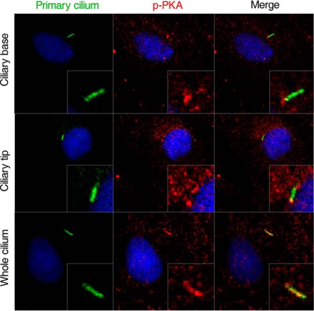
Figure 6.

Immunostaining localization of p-PKA in primary cilia of rat calvarial osteoblasts. Primary cilium are stained green (with acetylated α-tubulin), p-PKA stained red, and nuclei stained blue (with DAPI). Scale bar = 10 μm. Each experiment was conducted at least three times independently.
