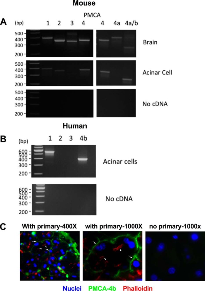
Figure 6.
PMCA isoforms are expressed in the pancreatic acinar cell. RNA was isolated from both mouse and human pancreatic acinar cells as well as whole brain as a positive control. First-strand cDNA was generated and used as a template for PCRs. Specific primers were used for the different PMCA isoforms. A, mouse. B, human. PMCA4b was also localized to the basal/lateral membrane of pancreatic acinar cells by immunofluorescence. C, representative image of pancreatic tissue labeled for PMCA4b (green), actin (phalloidin; red), and nuclei (DAPI; blue). Photomicrographs were taken at 400× or 1000× magnification. Arrowheads indicate apical actin staining, and arrows indicate predominately basal PMCA4b labeling.

