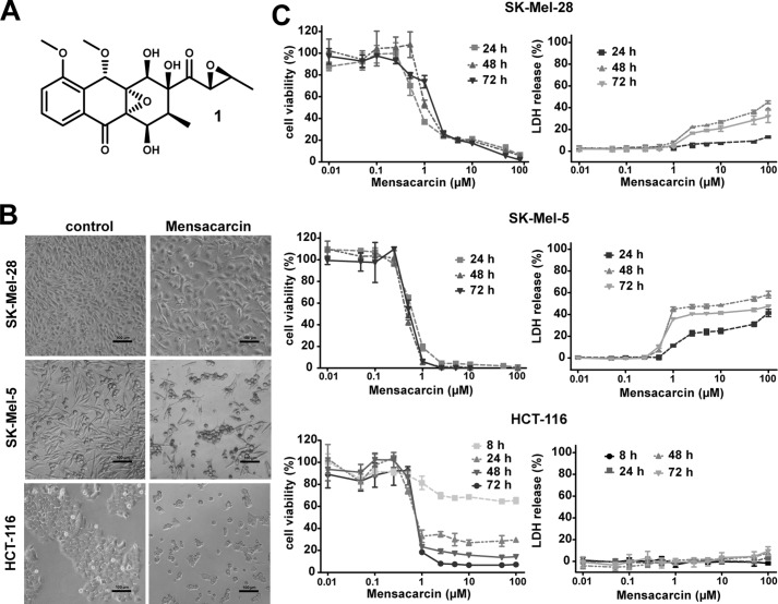
Figure 2.
Evaluation of cytostatic versus cytotoxic effects of mensacarcin in melanoma and colon cancer cells. A, chemical structure of mensacarcin (1). B, morphology of cells after 24-h exposure to 1 μm mensacarcin examined by phase-contrast microscopy. C, dose- and time-dependent growth inhibition and cytotoxicity of mensacarcin in SK-Mel-28 and SK-Mel-5 melanoma cells and HCT-116 colon cancer cells. Cell viability was determined by an MTT assay and LDH release assay as described under “Experimental Procedures.” Results are presented as mean ± S.D. (error bars) of three replicates (n = 3).

