Figure 3.
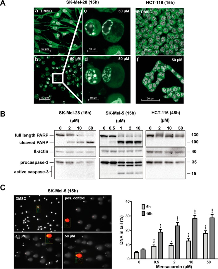
Mensacarcin induces rapid apoptotic cell death in melanoma cells. A, changes in nuclear morphology with chromatin condensation as a hallmark for apoptosis were examined by confocal fluorescence microscopy. Presented are images of SK-Mel-28 or HCT-116 cells that were grown in the presence of 0.5% (v/v) DMSO (a and e) or 50 μm mensacarcin (b and f) for 15 h and analyzed after staining with Hoechst 33342 and ER-Tracker Green. Insets show examples for cells with chromatin condensation in a single focal plane, which visualizes the necklace-shaped rings characteristic of apoptosis (c), or in a maximum intensity projection (d). B, apoptotic cell death was evaluated by immunoblot analysis of caspase 3 activation and PARP-1 cleavage. Shown are immunoblots of cell lysates of cells grown in the presence of various concentrations of mensacarcin or 0.25% (v/v) DMSO for 15 h (SK-Mel-28, SK-Mel-5) or 48 h (HCT-116). Analyzed lysates were pools of duplicates. β-Actin levels served as loading control. C, DNA damage associated with early apoptosis in SK-Mel-5 cells induced by mensacarcin was determined by an alkaline comet assay. Cells were grown in the presence of various amounts of mensacarcin or 0.5% (v/v) DMSO for 6 h or 15 h. At least 100 nuclei were randomly selected and scored for DNA damage. Results are presented as mean ± S.E. (error bars) (n = 100; two slides, 50 cells/slide). *, p < 0.1; ***, p < 0.001 versus DMSO control. B, representative images of mensacarcin-induced comets after 15-h treatment. Doxorubicin (5 μm) was used as a positive control.

