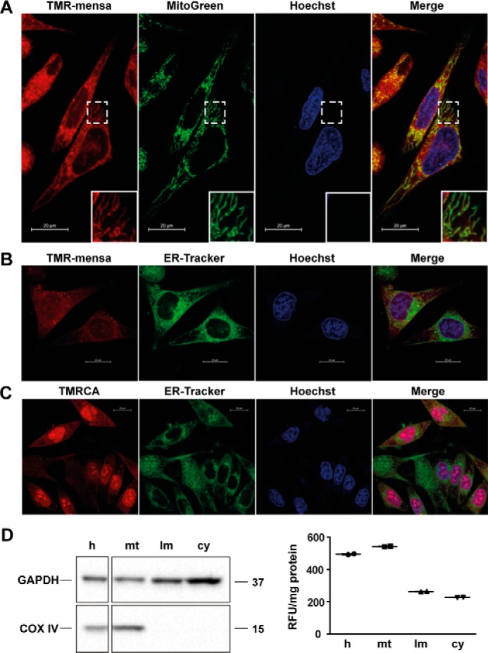
Figure 8.

Cell localization studies in melanoma cells with rhodamine-mensacarcin probe. A, SK-Mel-5 cells were stained with TMR-mensa, Hoechst 33342 (nucleus), and ER-Tracker Green (endoplasmic reticulum) or MitoGreen (mitochondria) for 20 min. Images were captured by inverted confocal fluorescence microscopy and merged to examine co-localization. TMR-mensa does co-localize with the MitoGreen with well-defined mitochondrial staining (inset). B, TMR-mensa does not co-localize with the ER-Tracker or Hoechst. C, control cells were stained with uncoupled rhodamine dye (TMRCA) and present an overall diffuse background fluorescence with dye accumulation in nuclei. D, cell subfractionation of TMR-mensacarcin–treated SK-Mel-5 cells (15-h exposure) into homogenate (h), mitochondrial fraction (mt), light membrane fraction (lm), and cytosolic fraction (cy). Fluorescence of each cellular subfraction was analyzed and normalized to protein content (excitation, 530 nm; emission, 590 nm). The enrichment of mitochondria in the mitochondrial fraction and the absence of mitochondria in the light membrane and cytosolic fractions was analyzed by immunoblot analysis using cytochrome c oxidase subunit IV (COX IV) as mitochondrial marker. The enrichment of cytosolic proteins in the cytosolic fraction was analyzed using GAPDH as marker protein.
