Figure 2.

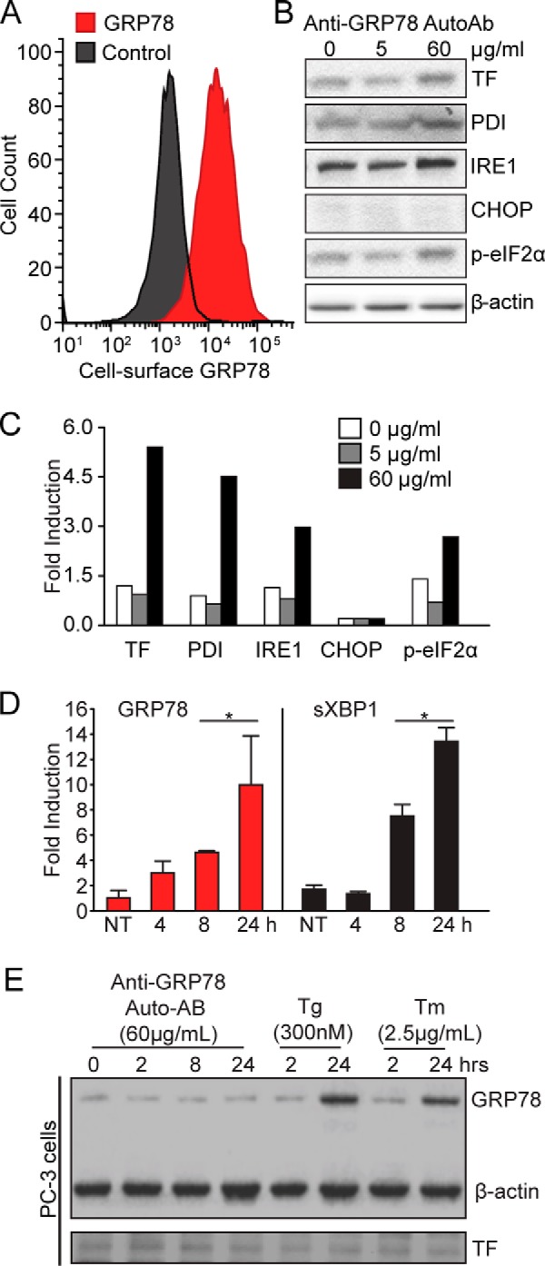
Treatment with anti-GRP78 AutoAbs increases the expression of TF and UPR markers in DU145 human prostate cancer cells. A, flow cytometry analysis of csGRP78 amounts in DU145 cells. B, pathological doses of anti-GRP78 AutoAbs (60 μg/ml) increase protein expression of both TF and markers of UPR activation (PDI, IRE1, and phospho-eIF2α), compared with non-treated (0 μg/ml) cells or cells treated with a normal dose of anti-GRP78 AutoAbs (5 μg/ml). β-Actin was used as a loading control. C, protein bands of the immunoblot in B were quantified with ImageJ, and values were normalized to β-actin. Error bars are ± S.D. p-eIF2α, phosphorylation of the initiation factor eIF2 α. D, quantitative real-time PCR analysis of GRP78 and spliced XBP1 mRNA expression in DU145 cells treated with a pathological dose of anti-GRP78 AutoAbs (60 μg/ml). Results are expressed as -fold induction over non-treated (NT) cells (*, p < 0.05; n = 3). E, pathological doses of anti-GRP78 AutoAbs (60 μg/ml) do not increase GRP78 expression in PC-3 cells; thapsigargin (Tg; 300 nm) or tunicamycin (Tm; 2.5 μg/ml) was used as a control UPR inducer. β-Actin was used as a loading control.
