Figure 4.
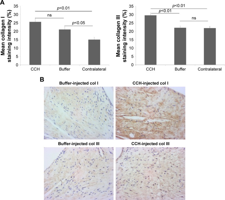
Immunostaining of collagen type I and collagen type III in the posterior capsule from contralateral knees and experimental knees intra-articularly injected with CCH or buffer.
Notes: (A) The extracellular matrix staining was quantified using ImageJ. Intensity of staining as a percentage is presented in the bar graphs (with total black considered as 100%). Experimental capsules (both CCH and buffer) showed significantly more staining than contralateral for collagen type I. Quantification of collagen type III staining for the same capsules showed a significant increase in staining in the CCH-treated capsule compared with buffer-treated and contralateral capsules (n=44 fields from buffer knees, n=36 fields from CCH knees, and n=64 fields from contralateral knees). (B) Representative micrographs of sections from the posterior knee capsule from joint injected with either buffer or collagenase (CCH) and stained with collagen type I or type III are shown.
Abbreviation: ns, not significant.

