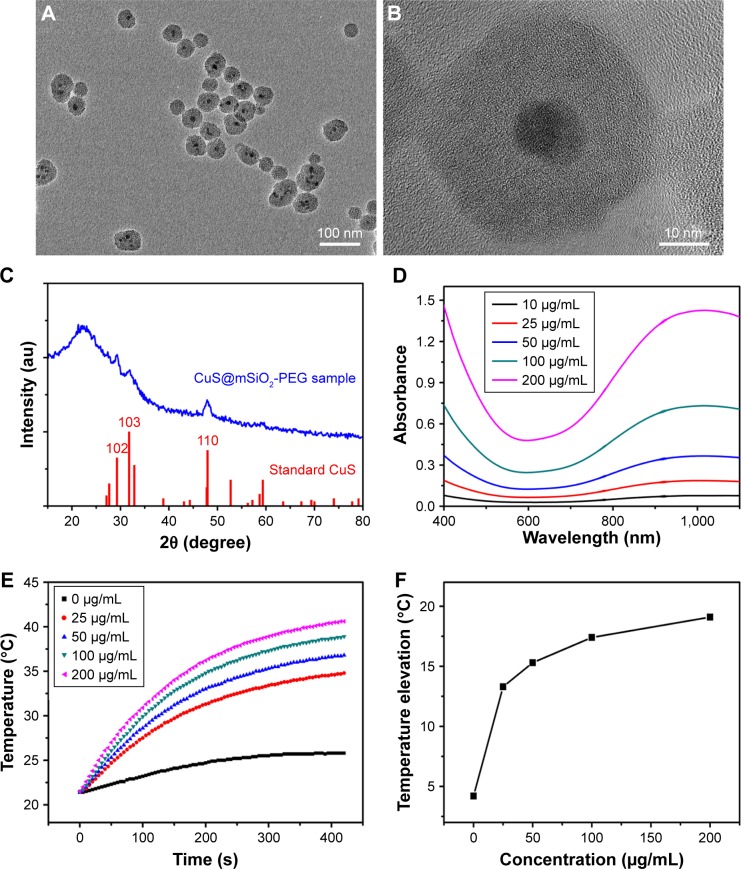
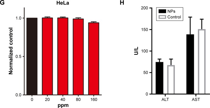
Figure 1.
Low (A) and high magnification (B) TEM image of the CuS@SiO2 NPs. XRD pattern (C) of the CuS@SiO2 NPs and the standard hexagonal phase of Cu (JCPDS card no: 06-0464). Absorption spectrum (D) for various concentrations of hydrophilic CuS@SiO2 NPs. Temperature elevation of pure water and of an aqueous dispersion of CuS@SiO2 NPs at different concentrations (0, 25, 50, 100, or 200 μg/mL) as a function of irradiation time (5 min). Pure water was used as a control, and the room temperature was 25°C (E). The temperature change (ΔT) over a period of 5 min has a significant positive correlation with the CuS@SiO2 NP concentration of the aqueous dispersion (F). The toxicity of the CuS@SiO2 NPs was evaluated by CCK-8 tests as a function of incubation concentration (0–160 ppm) for the HeLa cells (G). ALT and AST in blood biochemistry test (H). Cells were incubated with the solutions at 37°C for 24 h.
Abbreviations: CCK-8, Cell Counting Kit-8; NP, nanoparticle; TEM, transmission electron microscopy; XRD, X-ray diffractometer.


