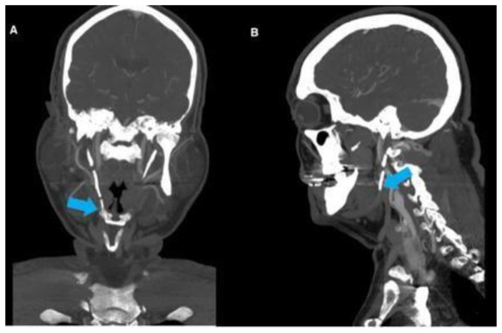
Figure 1.
69-year-old male with compression of the right ICA secondary to Eagle’s syndrome.
FINDINGS: Coronal (Fig. 1A) and Sagittal (Fig. 1B) contrast enhanced CT of the head and neck demonstrate post-endarterectomy appearance of the right ICA with an ipsilateral significantly elongated styloid process (arrows).
TECHNIQUE: Coronal (Fig. 1A) and Sagittal (Fig 1B) CTA, 3 mm slice thickness using Visipaque (320 mgl/ml) injected at a rate of 5 ml/sec for a total volume of 65 ml.

