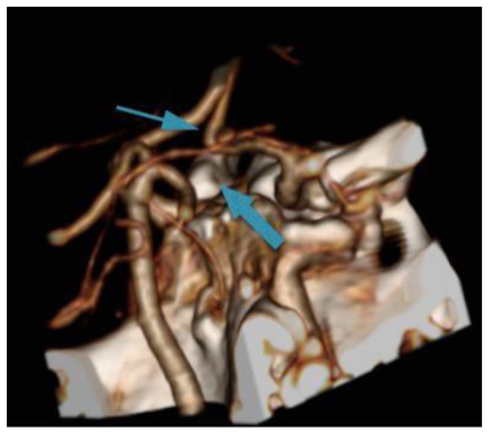
Figure 2.
69-year-old male with compression of the right ICA secondary to Eagle’s Syndrome.
FINDINGS: 3D CTA of the Circle of Willis demonstrates absent AComm (thin arrow) and absent PComm arteries (thick arrow). The arrows are pointing to the usual anatomical locations of both vessels.
TECHNIQUE: 3D reconstruction from a 3 mm slice thickness CTA using Visipaque (320 mgl/ml) injected at a rate of 5 ml/sec for a total volume of 65 ml

