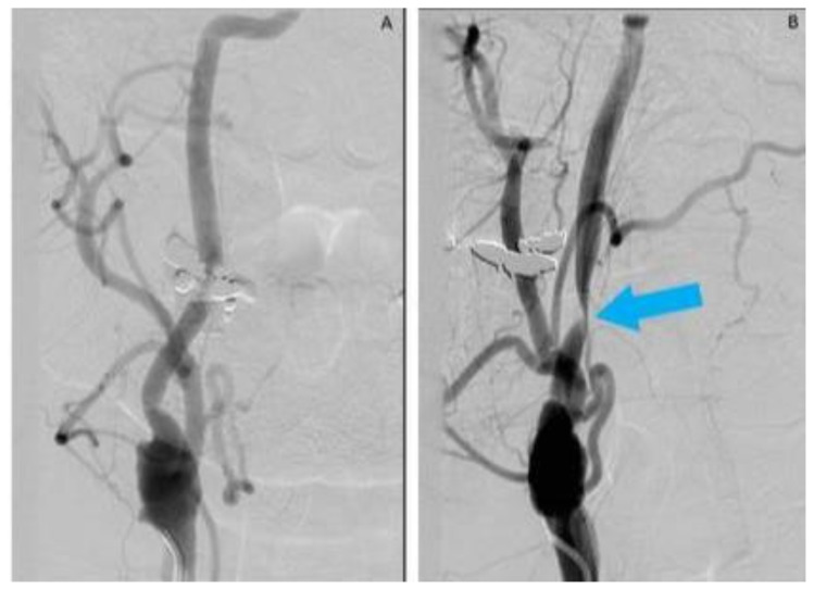
Figure 3.
69-year-old male with compression of the right ICA secondary to Eagle’s syndrome.
FINDINGS: Catheter angiogram in the arterial phase demonstrates normal filling in the post-endarectomy right ICA (Fig. 3A). When the patient turns his head to the right (Fig. 3B), severe right ICA stenosis is noted (arrow).
TECHNIQUE: Catheter angiogram of right common carotid artery using Omnipaque 300 (300mgl/ml) with the injection rate of 5 ml/sec for total volume of 12 ml.

