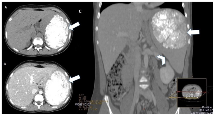
Figure 1.
15-years-old girl with beta-thalassemia, splenomegaly and a calcified splenic hamartoma. - Technique: Abdominal CT scans (120 kV, modulated mA, 3 mm slice thickness, 80 ml of Iomeron 400) - Findings: A: Axial non-contrast CT image shows a large (10 × 8 × 7 cm) splenic lesion (arrow) with many calcifications widely scattered inside. B: Axial contrast enhanced CT image on portal venous phase demonstrates the calcified non-enhancing splenoma (arrow), with well-defined margins. C: Coronal reformatted contrast enhanced CT of the abdomen (bone window) demonstrates the significantly enlarged spleen (arrowhead) with a calcified mass in the splenic parenchyma, related to splenoma (arrow).

