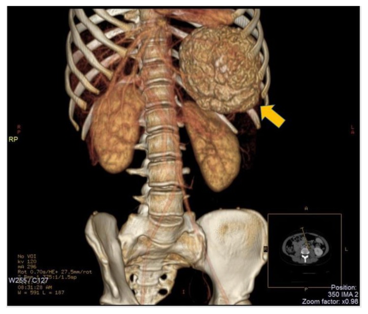
Figure 2.
15-years-old girl with beta-thalassemia, splenomegaly and a calcified splenic hamartoma. - Technique: Abdominal CT scan after intravenous administration of 80 ml of Iomeron 400 (120 kV, modulated mA, 3 mm slice thickness). 3D Volume Rendering of spiral CT data. - Findings: this image depicts the large splenic hamartoma (10 × 8 × 7 cm) with calcium density (arrow).

