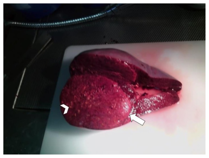
Figure 3.
15-years-old girl with beta-thalassemia, splenomegaly and a calcified splenic hamartoma. - Technique: standard macroscopy image after splenectomy - Findings: Gross photograph of the resected spleen illustrating a solid mass (arrow), measuring 10 × 8 × 7 cm, arising in the upper pole of the spleen (weighted 870 g). The lesion was well circumscribed from the surrounding splenic parenchyma but without a capsule and was dark red in appearance, mottled with numerous yellowish focal areas (arrowhead).

