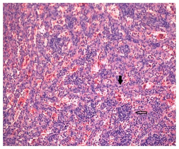
Figure 4.
15-years-old girl with beta-thalassemia, splenomegaly and a calcified splenic hamartoma. - Technique: standard hematoxylin-eosin stain, original magnification ×400 - Findings: High magnification of the lesion shows various lymphoid aggregates (empty arrow), interspersed with vascular structures (capillaries and sinusoids of splenic red pulp-like area) lined by endothelial cells with no cytologic atypia (black arrow).

