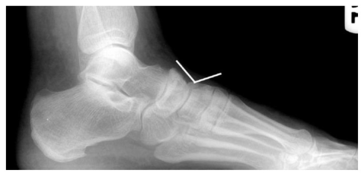
Figure 7.
This cropped lateral view of a 29 y/o male highlights the medial column of the foot with an associated “break” or “fault” in the navicular-cuneiform joint. One can appreciate a relative depression along the dorsal aspect of the bones (highlighted with the overlying white lines) at the level of the “fault” at the navicular-cuneiform joint.

