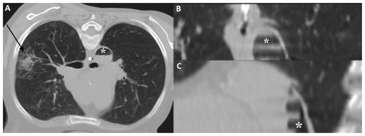
Figure 4.
57 year old female with systemic air embolism after percutaneous lung biopsy.
FINDINGS: Postprocedural axial CT scan (A) and reformatted images (coronal-B and sagittal-C) shows alveolar hemorrhage (black arrow) adjacent to the biopsied nodule and a significant amount of gas inside the thoracic aorta (asterisk), compatible with air embolism.
TECHNIQUE: Axial non-enhanced CT, 300mAs,120kV, 3mm slice thickness.

