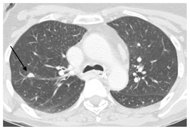
Figure 7.
57 year old female with systemic air embolism after percutaneous lung biopsy.
FINDINGS: axial CT scan performed two months and fourteen days after the procedure shows significant volumetric reduction of the lung nodule (black arrow). The patient was not under chemotherapy, suggesting a benign nature of the nodule.
TECHNIQUE: Axial non-enhanced CT, 300mAs,120kV, 3mm slice thickness.

