Figure 1.

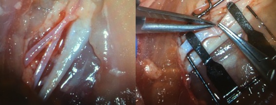
Photos showing the neurovascular structures of the chicken thigh model (left) and use of microvascular instrumentation to perform anastomosis (right).

Photos showing the neurovascular structures of the chicken thigh model (left) and use of microvascular instrumentation to perform anastomosis (right).