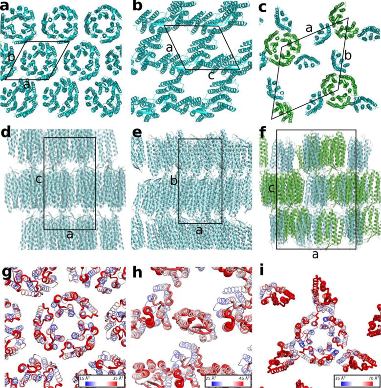
Figure 3.

Crystal packing of bR crystallized using monoolein, PDB ID 1M0L (a, d, g) and GlyNCOC15+4 as a host lipid for LCP at 4 °C (b, e, h) and 20 °C (c, f, i) as viewed
(a, b, c) in the membrane plane,
(d, e, f) along the membrane,
(g, h, i) in the membrane plane with B factors mapped as both thickness of the lines and as a gradient coloring with the blue color corresponding to the lowest values and the red color corresponding to the highest values.
The core bR trimer for the 20 °C structure is shown in green and the peripheral protomers are shown in cyan (panels c and f). Unit cells are outlined with solid black lines.
