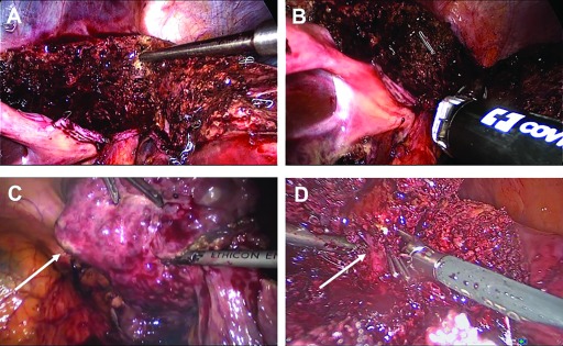
Figure 1.
Intraoperative photographs obtained during laparoscopic liver surgery. Parenchymal transection along the falciform ligament with the CUSA device for a left lateral sectorectomy (A). The hilar plate is dissected by endo-GIA staplers (B). Resection of segment 6 in a cirrhotic patient with HCC (arrow, C). Preparation of a major venous branch (arrow) during resection of a liver metastasis in segment 5/8 (D).

