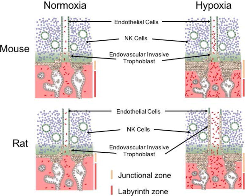
Figure 3. Maternal hypoxia stimulates adaptations at the mouse and rat placentation site.

Exposure of pregnant mice and rats to hypoxia (10–11% oxygen) during a critical developmental phase of development (gd 7.5 to 9.5) results in striking changes in the organization of hemochorial placentation. In both species, there is an increased allocation of trophoblast progenitor cells to differentiation of junctional zone trophoblast cell lineages instead of labyrinth zone trophoblast cell lineages. In the rat but not the mouse, maternal hypoxia also activates development of endovascular trophoblast cell lineage, and accelerates endovascular trophoblast cell invasion, which restructure uterine spiral arterioles. The schematic presentation is based on previously reported findings (Rosario et al., 2008, Bu et al., 2016).
