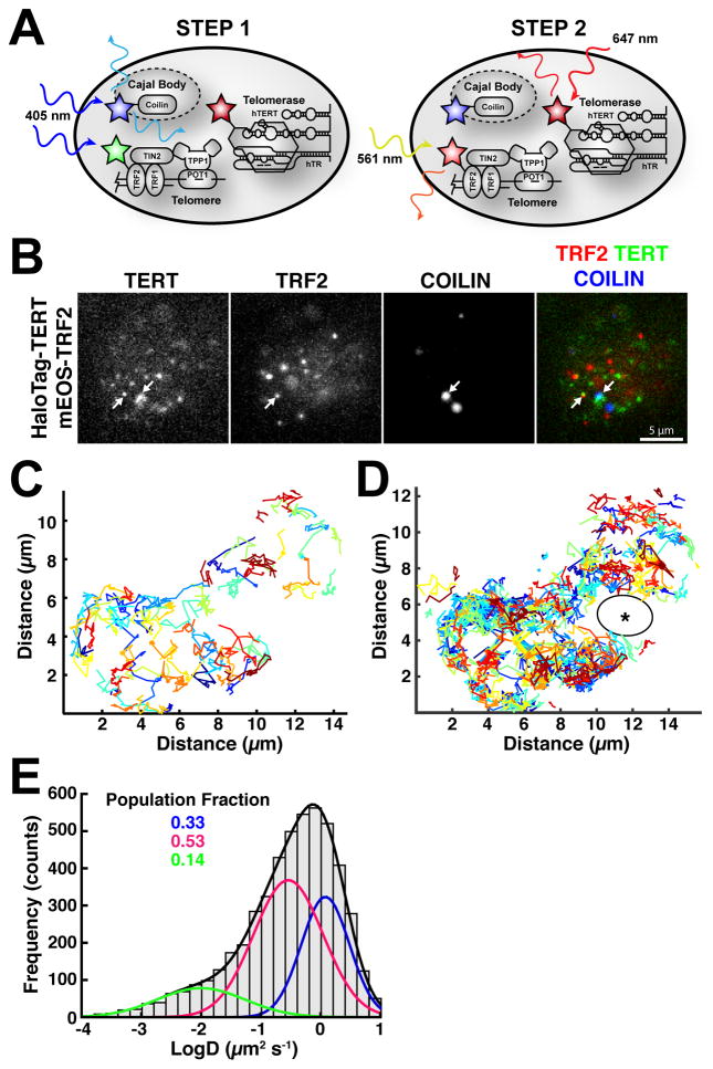
Figure 2. Three-dimensional Diffusion of Telomerase Governs its Search for Telomeres.
(A) Diagram illustrating our approach for visualizing Cajal bodies, telomerase, and telomeres. First, BFP-coilin was imaged, also converting mEOS-3.2 from the green to the red state. Immediately afterwards, we simultaneously imaged red mEOS3.2-TRF2 and HaloTag(JF646)-TERT at 45 frames per second. (B) Still images from movies simultaneously visualizing telomeres (mEOS3.2) and telomerase (HaloTag-JF646), after imaging Cajal bodies marked by BFP-coilin (white arrows indicate co-localizations). (C) A subset of trajectories of telomerase particles, present for at least 10 frames (~220 ms), generated by single-particle tracking of telomerase signals at 45 fps (20 ms exposure), demonstrating rapid three-dimensional diffusion. (D) All TERT trajectories detected in a 45 s movie. Unexplored region marked by asterisk. (E) Diffusion coefficient histogram of telomerase tracks present for at least 5 consecutive frames (N = 18 cells, n = 5035 tracks). Two freely diffusing populations (D ~ 0.3 μm2/s, magenta; D ~ 1.3 μm2/s, blue) and a smaller less mobile population (D ~ 0.01 μm2/s, green) are present. Fractions of the total number of particles in each population are indicated. Also see Movies S1–S4.

