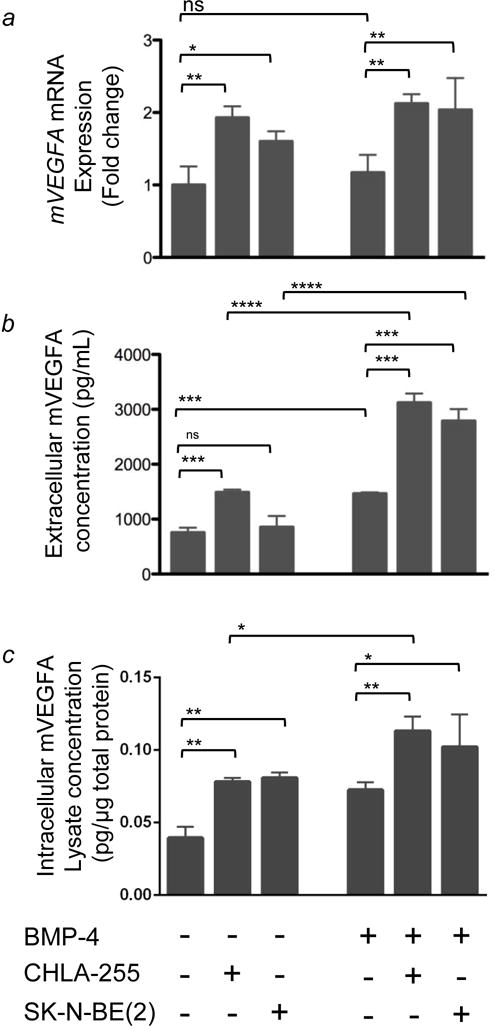
Figure 3. NBL cells induce an increase in mVEGFA expression in primary mBMMSC.

BMMSC were co-cultured in the presence or absence of human NBL cells as described in Figure 1. (a) mVegfa mRNA expression in BMMSC was quantified by qRT-PCR. Columns 5 and 6 show a 2 fold increase in mVEGFA in BMMSC cultured in the presence CHLA-255+BMP-4 and SKNBE(2)+BMP-4, respectively, compared to BMMSC cultured in the absence of NBL cells. The data represent the mean fold change (±SD) of gene expression from triplicate samples (n=3) and represent one of two experiments showing similar results. (b) Extracellular mVEGFA protein levels in the medium of BMMSC cultured in conditions indicated above. The data represent the mean protein level (±SD) of triplicate samples and is representative of similar results from 3 experiments. (c) Intracellular mVEGFA protein levels in BMMSC lysate expressed as pg/μg of total protein. Data is from triplicate samples from one experiment. In all panels, * = p<0.05, ** = p<0.01, *** = p<0.001, **** = p<0.0001
