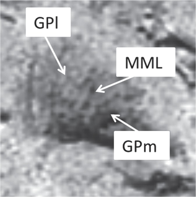
Fig 5.

Examples of the grading of the MML on PADRE images from NC, a 70-year-old man, taken on a coronal plane. Although elderly subjects also often showed a good depiction of MML, whole MML was not delineated (grade 3). MML, medial medullary lamina; GPm, medial globus pallidus; PADRE, phase difference-enhanced; GPl, lateral globus pallidus; NC, normal control subjects.
