Figure 1.

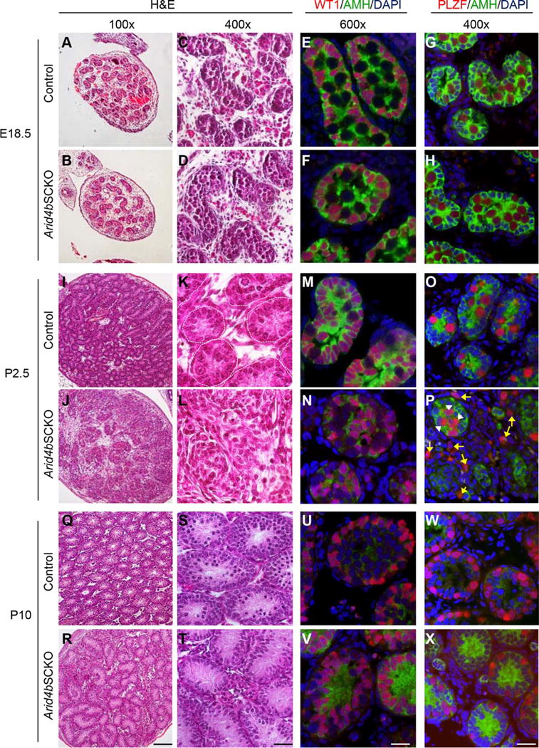
Failure to establish the spermatogonial stem cell niche in the Arid4bSCKO testes at P2.5 of age. (A–D, I–L, Q–T): Histological analyses of the control and Arid4bSCKO testes at E18.5, P2.5, and P10. Paraffin-embedded testis sections were stained with H&E. The basement membrane of the seminiferous tubules is outlined with dashed lines (K). Original magnifications of images were 100× (A, B, I, J, Q, R) and 400× (C, D, K, L, S, T). Scale bars = 100 μm (R) and 25 μm (T). (E, F, M, N, U, V): Double immunofluorescent staining of anti-Müllerian hormone (AMH) (green, cytoplasmic) and Wilms Tumor 1 (red, nuclear) to detect Sertoli cells in testis sections from the Arid4bSCKO and control mice at E18.5, P2.5, and P10. DNA was stained by DAPI (blue). Scale bar = 20 μm. (G, H, O, P, W, X): Double immunofluorescent staining of AMH (green, cytoplasmic) and promyelocytic leukemia zinc finger (red, nuclear) to detect Sertoli cells and gonocytes, respectively. Testis sections were from the Arid4bSCKO and control mice at E18.5, P2.5, and P10. Nuclear DNA was stained by DAPI (blue). White arrowheads point to gonocytes at central location within the seminiferous cords, and yellow arrows point to gonocytes scattered outside the cords in the Arid4bSCKO testes at P2.5 (P). Scale bar = 25 μm. Abbreviations: AMH, anti-Müllerian hormone; DAPI, 4′,6-diamidino-2-phenylindole; PLZF, promyelocytic leukemia zinc finger; WT1, Wilms Tumor 1.
