Figure 9.
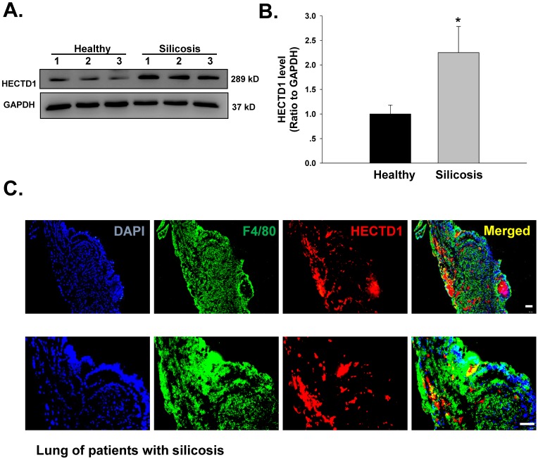
HECTD1 expression is increased in macrophages from patients with silicosis. (A) Representative western blot showing HECTD1 expression in macrophages from healthy donors and patients with silicosis. (B) Densitometric analyses of macrophage samples from five healthy donors and five patients with silicosis suggested that HECTD1 expression was elevated in macrophages from patients with silicosis. *P<0.05 vs. the corresponding healthy control group. (C) Immunohistochemistry of the macrophage marker F4/80 and HECTD1 in lung tissues from patients with silicosis. Colocalization between F4/80 and HECTD1 is shown. The images are representative of several individuals from each group (n=5). Scale bar=20 μm.

