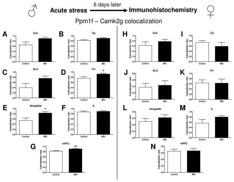
Figure 5. Stress immobilization and Ppm1f/Camk2g levels in the Amygdala and mPFC in both male and female mice: an immunohistochemistry study.
Quantification of Ppm1f and Camk2g levels reveals that both are more densely colocalized after traumatic stress in male mice in the Amygdala and mPFC. (A,H) CeA = Central amygdala, (B,I) Cg = Cingulate cortex, (C,J) BLA = Basolateral amygdala, (D,K) Prl= Prelimbic cortex, (E,L) (Total) Amygdala = CeA + BLA, (F,M) IL= Infralimbic cortex, (G,N) mPFC = Medial prefrontal cortex = Cg+Prl+IL. *p≤0.05, **p≤0.01. N=2 per group.

