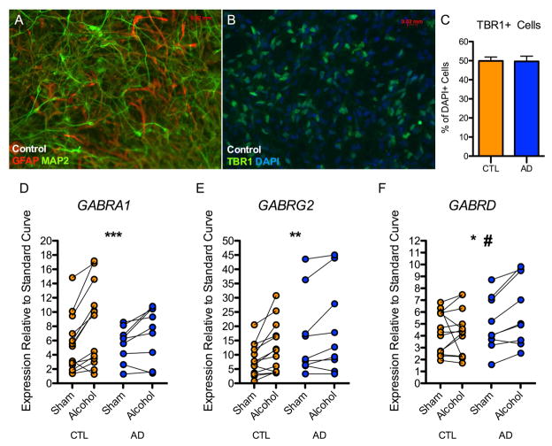
Figure 1. Effect of 21-day alcohol exposure on GABAA subunit mRNA expression.
12–14 week old neural cultures derived from controls and alcoholics were characterized by immunocytochemistry. (A) Representative image from a control subject indicating iPSCs differentiate into mixed neural cultures containing MAP2-postive neurons and GFAP-positive astrocytes. (B) Representative image from a control subject indicating that cultures are enriched for TBR1+ forebrain-type glutamate neurons. (C) No difference was observed in the number of TBR1+ neurons between neural cultures derived from controls and alcoholics. 10,255 DAPI+ cells derived from 6 control and 6 alcoholic neural cell lines were analyzed. (D, E, F) 12-week old neural cell lines (13 control and 9 alcoholic) were treated daily with neural media supplemented with 50 mM alcohol. (D) A significant effect of alcohol treatment was observed for GABRA1 gene expression. (E) A significant effect of alcohol treatment was observed for GABRG2 gene expression. (F) A significant effect of alcohol treatment was observed for GABRD gene expression. A modestly significant interaction between donor status (alcoholic vs. control) and alcohol treatment was observed. (Two-way repeated measures ANOVA: *p < 0.05, **p < 0.01, ***p < 0.001 for alcohol treatment; #p = 0.054 for interaction between donor status and alcohol treatment)

