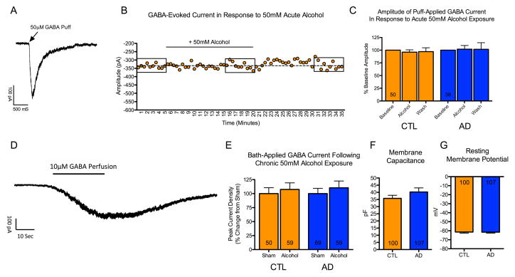
Figure 2. Effect of acute and chronic alcohol on GABA-evoked currents.
Mature neurons (22–36 weeks post plating) derived from 6 control and 7 alcoholic lines were used for electrophysiological recordings. (A) Example trace of a GABA-evoked current in response to brief pressure application of 50 μM GABA. (B) Example recording of a neuron from a control subject. Each dot represents a GABA-evoked response, which was evoked every 30-seconds. Following a 5-minute baseline recording, aCSF containing 50 mM alcohol was perfused for a total of 15 minutes, which was subsequently washed out for an additional 15 minutes. Boxes indicate responses within 5-minute bins that were averaged for analysis. (C) No significant effect of acute alcohol was observed on the amplitude of GABA-evoked responses in neurons derived from 6 control or 7 alcoholic iPSC lines. Washout contains data from a subset of 46 control neurons and 32 alcoholic neurons. (D) Example trace of a GABA response in a control neuron evoked by bath perfusion of aCSF supplemented with 10 μM GABA. Bath perfusion of GABA evoked a reversible current that plateaued after 30–40 seconds. (E) No difference in maximum current density of GABA-evoked current was observed following 9–21 days of exposure to 50 mM alcohol in neurons derived from 4 control or 5 alcoholic iPSC lines. (F and G) No significant difference was observed in membrane capacitance (an indicator of cell size) or resting membrane potential in non-alcohol treated neurons derived from the 6 control and 7 alcoholic iPSC lines. (Numbers within bars indicate the total patched neurons).

