Figure 2.
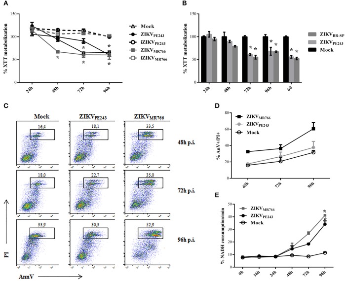
Cell viability of HBMECs infected with ZIKV. (A) HBMECs were mock-treated or cultured with ZIKVPE243 or ZIKVMR766, produced in C6/36 cells, in their native and inactivated forms (iZIKV). XTT metabolization was assayed at the indicated time points, and normalized according to the values obtained in cell cultures maintained in culture medium only. Data are represented as mean ± SD of six independent experiments. *p < 0.05 in relation to mock. (B) HBMECs were mock-treated or infected with ZIKVPE243 or ZIKVBR−SP and XTT assay was performed as in (A). (C,D) HBMECs were infected with ZIKVPE243 or ZIKVMR766. Cells were stained with AlexaFluor488-Annexin V (AnnV) and propidium iodide (PI) and were evaluated by flow cytometry, at the indicated time points. (C) Dot plot indicating the percentage of AnV+PI+ cells at each time point, in a representative experiment. (D) Average percentage of AnnV+PI+cells at each time point from three independent experiments. (E) Culture supernatants were harvested and LDH release was evaluated by measurement of NADH consumption/min in a LDH activity assay. Data are represented as mean ± SD of three independent experiments. *p < 0.05 in relation to mock.

