Figure 7.
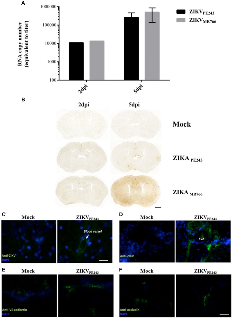
ZIKV reaches mouse brains without disrupting BBB. A129 mice were mock-inoculated or infected with ZIKVPE243 or ZIKVMR766 (2 × 10⁵ PFU) by i.v. route. (A) Virus RNA in the brains obtained from 2 or 5 days infected mice were measured by qRT-PCR. The values indicates the average of RNA copy numbers of four individual mouse infected with the respective viral strain. (B) Photomicrographs showing the pattern of immunoglobulin G staining in the brain of young A129 mice infected with ZIKVMR766 or ZIKVPE243, at 2 or 5 days post infection (dpi). Mice from the control group received an intravenous injection of saline. Scale bar: 1,000 μm. (C–F) After 5dpi, mice were transcardially perfused, the brains were harvested, and immunohistochemistry analyses were performed as described. Brain sections were incubated with mouse antibodies anti-4G2 antibody (C,D), anti-VE-cadherin (E), or anti-occludin (F), followed by incubation with AlexaFluor 488-conjugated anti-mouse IgG. Expression of virus E protein and adherens and tight junction were then analyzed using a Zeiss Axio Observer Z1 microscope equipped with an Apotome module.

