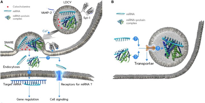
Figure 1.
Schematic diagram of the microRNAs (miRNA) exocytosis mechanisms (A) and the working hypothesis of the miRNA loading into large dense-core vesicles (LDCVs) (B). (A) Catecholamines (red ball) are typical neurotransmitters stored in LDCVs. LDCVs also contain a variety of miRNAs including miR-375. The assembly of neuronal SNAREs including VAMP-2, SNAP-25A, and syntaxin-1A mediates miRNA exocytosis from chromaffin cells, neuroendocrine cells. Synaptotagmin-1 (Syt-1) is considered as a Ca2+ (green ball) sensor to trigger miRNA exocytosis. The membrane insertion of Ca2+-bound Syt-1 results in the fusion pore formation. Ribomone hypothesis: miRNAs stored in vesicles together with classical neurotransmitters are released by vesicle fusion, thereby contributing to cell-to-cell communication (24). Two hypothetical functions of released extracellular miRNAs; (i) miRNAs might be taken up by endocytosis into target cells where miRNAs regulate gene expression. (ii) miRNAs might be able to stimulate receptors or ion channels as ligands, thereby leading to cellular signalling. Adapted from Gümürdü et al. (24). (B) The mechanisms by which miRNA or miRNA–protein complex can be loaded into LDCVs remain elusive. Structure of miRNA-binding protein is artificial for the simplicity.

