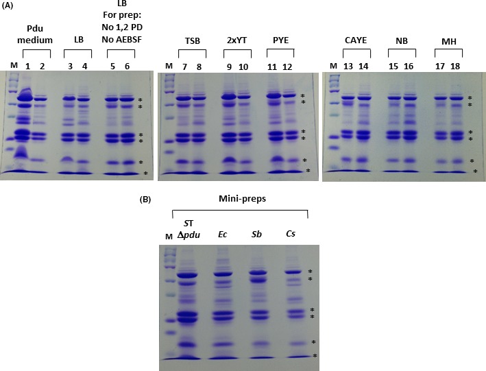
Figure 5.

Analysis of R995 + pdu ST MCP isolation using different growth media and a mini‐prep protocol. Panel A: S. bongori and C. sakazakii strains containing R995 + pdu ST were grown in the indicated media supplemented with 1,2 PD, and the MCPs were isolated from each culture. An aliquot of each MCP prep (corresponding to approximately 10–20 μg protein) was run via SDS‐PAGE and stained with Coomassie Blue. The ‘M’ lane is protein marker (15 μg), odd numbered lanes contain samples from S. bongori R995 + pdu ST, and even numbered lanes contain samples from C. sakazakii R995 + pdu ST. Abbreviations correspond to those indicated in the Experimental Procedures for each medium. Control strains containing the R995 vector did not display MCP bands when grown and analysed in these media (data not shown). The asterisks on the right side of each gel indicate bands corresponding to known S. Typhimurium Pdu proteins. The sample ‘LB, For prep: no 1,2 PD, no AEBSF’ indicates MCP preps obtained from cells grown in LB medium (containing 1,2 PD) but with no 1,2 PD or protease inhibitor present during the purification procedure. Panel B: The indicated strains containing R995 + pdu ST were grown in LB medium supplemented with 1,2 PD, and the MCPs from each culture were isolated using a mini‐prep protocol as described in Experimental Procedures. An aliquot of each MCP mini‐prep was run via SDS‐PAGE and stained with Coomassie Blue. Species abbreviations correspond to those indicated in the legend for Fig. 1.
