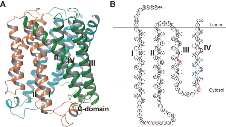
Figure 2.
Structure of mPGES-1. (A) Overall structure of the human PGES-1 trimer (PDB4AL0).19) (B) Schematic model structure of the human mPGES-1 monomer. The red letters show the residues critical for mPGES-1 enzymatic activity,14,20) and the blue letters show the residues that account for the species discrepancy in the human-specific mPGES-1 inhibitor.57) I, II, III, and IV indicate transmembrane helix 1, 2, 3, and 4, respectively.

