
FIGURE 3.
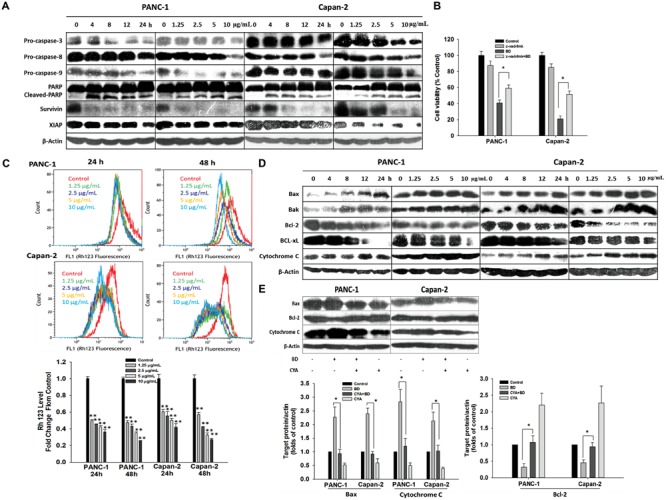
BD triggers caspases/mitochondria-dependent apoptosis. (A) Cells were treated with BD either at concentrations of 1.25, 2.5, 5, and 10 μg/mL for 12 h or at 5 μg/mL for various durations of 4, 8, 12, and 24 h. The cell lysates harvested were analyzed for expression of PARP-1, pro-caspase-3, pro-caspase-8, pro-caspase-9, Survivin and XIAP by Western blotting. β-Actin served as the protein loading control. (B) Z-VAD-FMK (caspase inhibitor, 50 μM) was added simultaneously with BD to PANC-1 and Capan-2 cells. Cells were harvested for cell viability analysis by MTT assay. (C) Cells were treated with BD at the indicated concentrations for 24 and 48 h and were stained by Rhodamine 123 to determine the change of ΔΨm by flow cytometry. (D) Protein expression involved in the mitochondrial function were measured by Western blotting in both PANC-1 and Capan-2 cells treated with various concentrations of BD for 12 h or at 5 μg/mL for different treatment durations. β-Actin served as the protein loading control. (E) Western blotting analysis of Bax, Bcl-2, and cytochrome c levels after cells were incubated with cyclosporine A (10 μM) or BD (5 μg/mL) alone or in combination for 24 h. β-Actin served as the protein loading control. Each bar represents means ± SD of three independent experiments. ∗P < 0.05 and ∗∗P < 0.01 vs the control group.