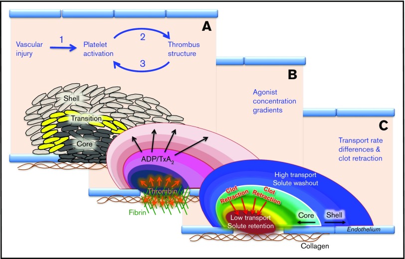
Figure 1.
A systems view of the hemostatic response to penetrating injuries. High-resolution confocal fluorescence microscopy studies performed in mice show that the hemostatic structure has a characteristic architecture whose properties emerge as platelets accumulate, altering local conditions. (A) Presence of a core of highly activated, densely packed platelets overlaid with a shell of loosely packed, less activated platelets and the transition zone that exists between them. (B) Manner in which soluble platelet agonists such as thrombin, ADP, and TxA2 form concentration gradients radiating from the site of injury. (C) Distribution of the agonists is determined in part by differences in transport rates in the narrowing gaps between platelets, gaps whose dimensions decrease as clot retraction proceeds.

