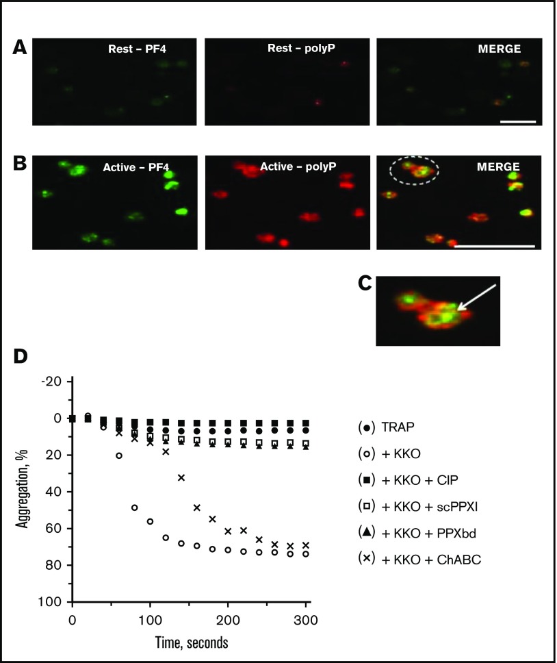
Figure 6.
Subcellular localization of PF4 and polyP in resting and stimulated platelets. Human platelets were plated onto glass coverslips in the resting state (A, top) or after <1 minute of activation with 100 nM of the phorbol ester, PMA, and 1 μM of calcium ionophore A23187 (B, middle). After immediate fixation and permeabilization, the platelets were stained to detect PF4 (left, green) and polyP (middle, red) by confocal microscopy. The panels on the right reveal the merged images (yellow). In resting platelets, PF4 and polyP are clearly contained primarily within separate granules (A). After activation, PF4 and polyP coalesce toward the center of the platelets where they colocalize (B). Bars represent 10 μm. C, Higher magnification of the area indicated by the dotted circle in panel B, right panel, depicting centralization and colocalization of PF4 and polyP (arrow). (D) Washed platelets were incubated with chondroitinase ABC, CIP (20 or 200 U/mL), PPX1 (2-10 µg/mL), or PPXbd (10-125 µg/mL), followed immediately by addition of 1 µM TRAP ± 100 µg KKO. Platelet aggregation was followed over the ensuing 250 seconds. The data shown are representative of results of 3 separate experiments.

