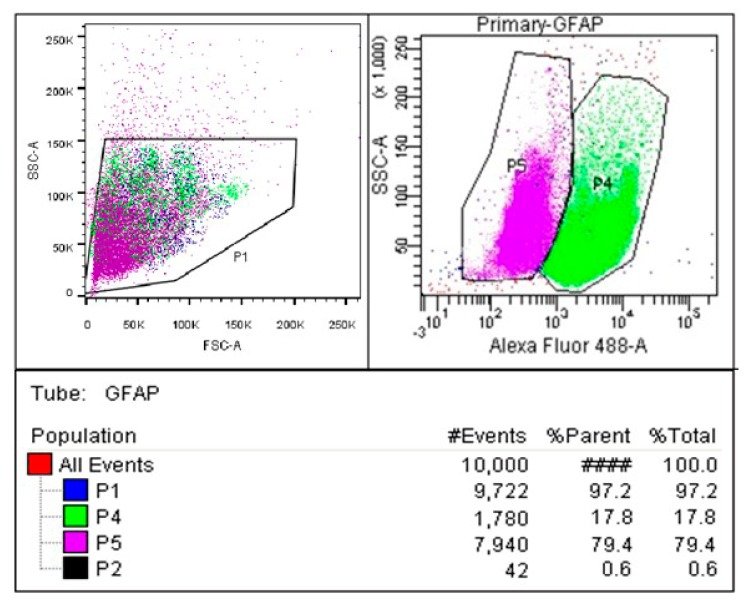
Figure 1.
Glial fibrillary acidic protein (GFAP) immunophenotyping to determine the purity of astrocyte culture. Cultures were stained with GFAP antibodies tagged with FITC (Fluorescein Isothiocyanate) and analyzed on a flow cytometer. P1 shows the population of cells; P2 shows the debris cells which are located in the lower left-hand side of scatter plots; P4 shows the primary astrocyte cells; P5 shows the contaminated cells. The plot shows that 79.4% of cells in the culture are GFAP+.

