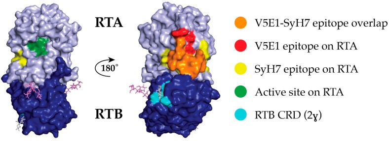
Figure 1.
V5E1 and SyH7 epitope localized on surface depiction of ricin toxin. Ricin holotoxin (PDB ID 2AAI) displayed in PyMol showing ricin’s enzymatic subunit (RTA; light blue) and ricin toxin’s binding subunit (RTB; dark blue). The overlap between SyH7 and V5E1 epitopes is shown in orange; SyH7’s additional epitope coverage is in yellow, and V5E1’s additional epitope coverage is in red. Also highlighted are RTA’s active site (green), and RTB’s 2γ Gal/GalNAc binding pocket (sky blue) with lactose molecule (gray) (right image).

