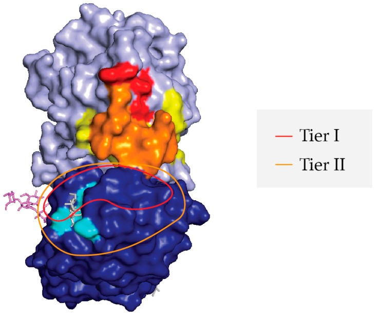
Figure 7.
Proposed Supercluster II toxin-neutralizing hotspot at the RTA-RTB interface. PyMol image of ricin holotoxin (as per Figure 1) with the following landmarks highlighted: RTA (light blue), RTB (dark blue), RTB’s 2γ Gal/GalNAc binding pocket (aqua blue), SyH7’s epitope (yellow), V5E1’s epitope (red), and the overlap of SyH7 and V5E1 epitopes (orange). The Tier I VHH epitopes are proposed to be confined within the red trace, while the Tier II VHH epitopes are proposed to be within the orange trace.

