Figure 1.

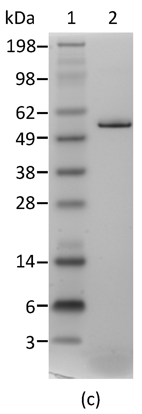
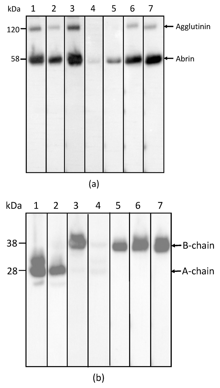
Reactivity of mAbs to abrin and agglutinin determined by Western blot analysis. Each lane was loaded with 250 ng of the abrin preparation and separated by SDS-PAGE under non-reducing (a) and reducing (b) conditions. Membranes 1–7 were incubated with mAbs, Abrin-1, to -7, respectively. The sizes of the agglutinin and abrin are indicated in kilodaltons (kDa) at the left side of panel (a) and the sizes of the A-chain and B-chain of the abrin/agglutinin are indicated at the left side of panel (b). The purity of the abrin (1 µg) used in this study was examined by non-reducing SDS-PAGE, followed with SimpleBlue staining (c).

