Figure 1.
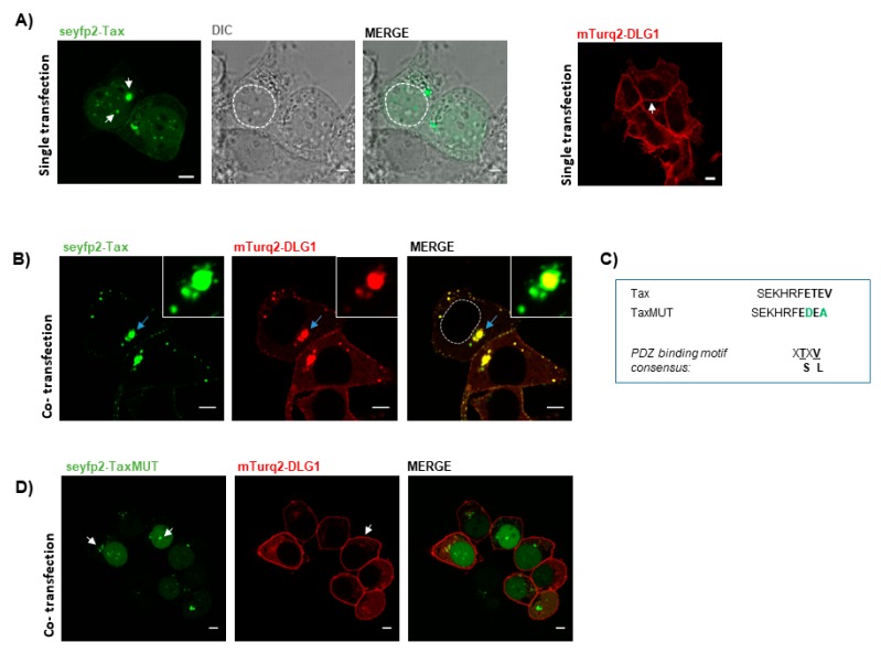
Simultaneous overexpression of discs large homolog 1 (DLG1) and human T cell leukemia virus (HTLV)-1 Tax (Tax) proteins generates a well-defined co-localization pattern. (A) Subcellular distribution of DLG1 and Tax proteins. Human Embryonic Kidney 293 (HEK293) cells transiently transfected with pseyfp2-Tax (green, left panel) or pmTurq2 -DLG1 (red, right panel) were fixed and protein expression was examined by confocal microscopy. The differential interference contrast (DIC) image is shown for better interpretation of nuclear localization; (B) Co-localization analysis of DLG1 and Tax. The pseyfp2-Tax and pmTurq2-DLG1 plasmids were simultaneously transfected into HEK293 cells and DLG1-Tax co-localization was assessed. The dashed circle delineates the nuclear region; (C) C-terminal sequence of Tax. The PBM of the Tax protein is highlighted with bold letters. Amino acid substitutions (green letters) introduced into the Tax PBM are shown with respect to the wild type Tax sequence. The consensus PBM sequence is also shown; (D) Co-localization analysis of DLG1 and Tax C-terminal mutant derivative (TaxMUT). The pseyfp2-TaxMUT (green) was transfected in HEK293 cells along with the pmTurq2-DLG1 plasmid (red). The co-localization between the proteins was assessed by confocal microscopy. In (A,D), the white arrows show the particular expression of each protein as described in the text. In (B), the light blue arrows indicate the co-localization region amplified in the inset. Dashed circles indicate nuclear regions. All scale bars represent 5 µm.

