
Figure 4. Retinoblastomas originate in the retina.
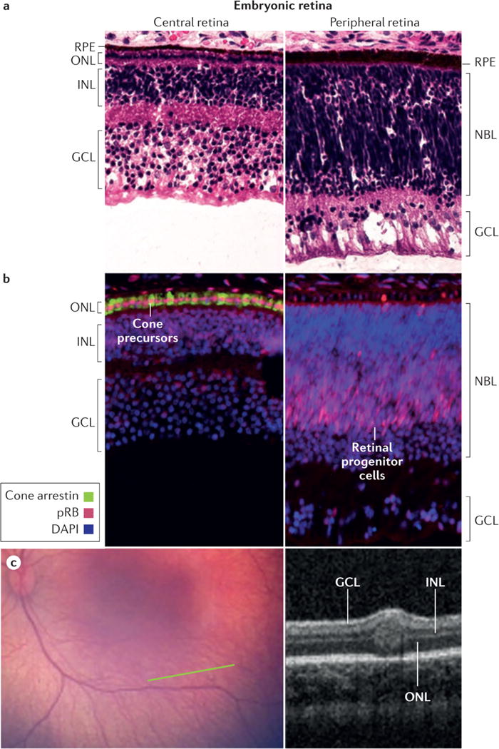
a, The retina has a complex structure and contains multiple cell types. Haematoxylin and eosin staining of post-fertilization week 19 retina shows three post-mitotic nuclear layers in the central retina near the fovea (left) and two nuclear layers in the less mature periphery of the same histologic section (right). Cell types in each layer are indicated. b, Cone precursors normally have high expression of the RB protein (pRB). Immunofluorescence staining of post-fertilization week 19 retina shows especially strong pRB signal (pink) in maturing cone precursors in the central retina (left, counterstained for cone arrestin, green) and in retinal progenitor cells in the peripheral retina of the same histologic section (right). pRB staining is less intense in DAPI-stained nuclei (blue) in other retinal cell types. c, Wide-angle retinal image (left image) shows no tumor. In the plane of the green line, optical coherence tomography (OCT; right image) shows a tiny intra-retinal tumor in a 2.5 month old infant, which seems to extend from the inner nuclear layer to the outer nuclear layer. The tumour has an uncertain epicentre making it difficult to infer the retinal layer from which the tumor arose. The layer-of-origin may be defined in the future with more images of higher resolution OCT of retina in very young children carrying an inherited germline RB1 mutation. Abbreviations: ONL, outer nuclear layer; INL, inner nuclear layer; GCL, ganglion cell layer; NBL, neuroblastic layer; RPE, retinal pigment epithelium.