
Figure 5. Online diagnosis of retinoblastoma.
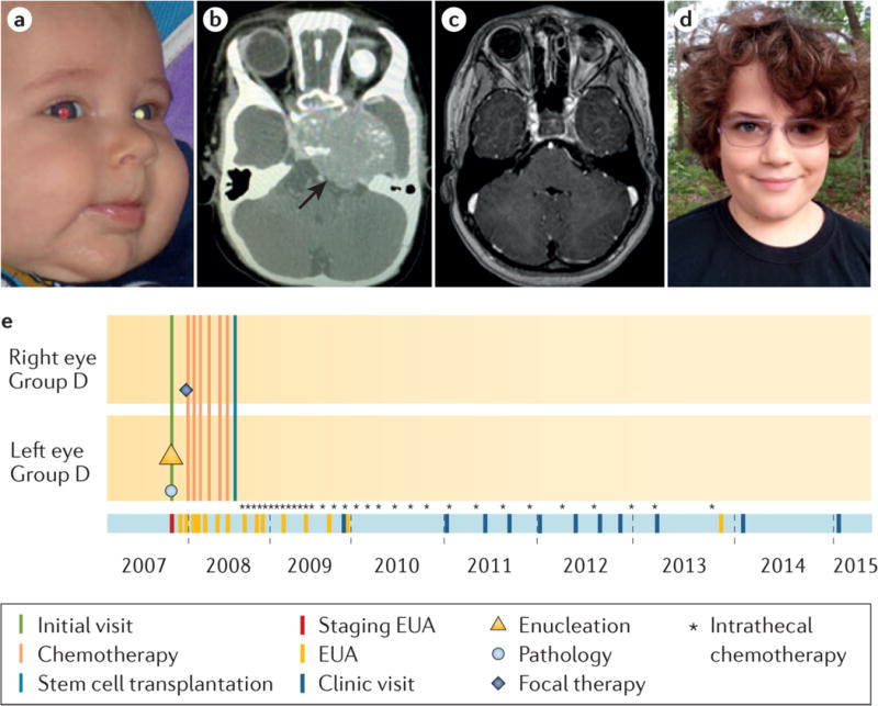
a, Detection of photoleukocoria (white pupil) on this digital image led the parents to the diagnosis of retinoblastoma. The left eye was determined by examination under anaesthetic to have IIRC78 Group D retinoblastoma and was enucleated two days later; CT scan was scheduled to check for trilateral tumour. (Today, MRI would be recommended to reduce radiation exposure.) The pathologic examination of the eye showed no high-risk features. The other eye was normal at diagnosis but on the next examination a small tumour was detected and treated with only laser. b, One month after surgery, the child presented with high intracranial pressure. A large intracranial midline tumour (trilateral disease) was diagnosed on CT scan, which was treated with chemotherapy, high-dose chemotherapy with hematopoietic rescue by stem cell transplant, and intrathecal chemotherapy injections (directly into the cerebro-spinal fluid) through an implanted intraventricular catheter. c, Follow-up MRI at age 8 years shows no residual disease and d, the child is well with one good-looking artificial eye and one eye with normal vision; he wears poly-carbonate lenses to protect his only eye. e, The eCCRB timeline of treatments; not all intrathecal chemotherapy injections (*) are shown.