
Figure 9. Retinoblastoma treated with IVC.
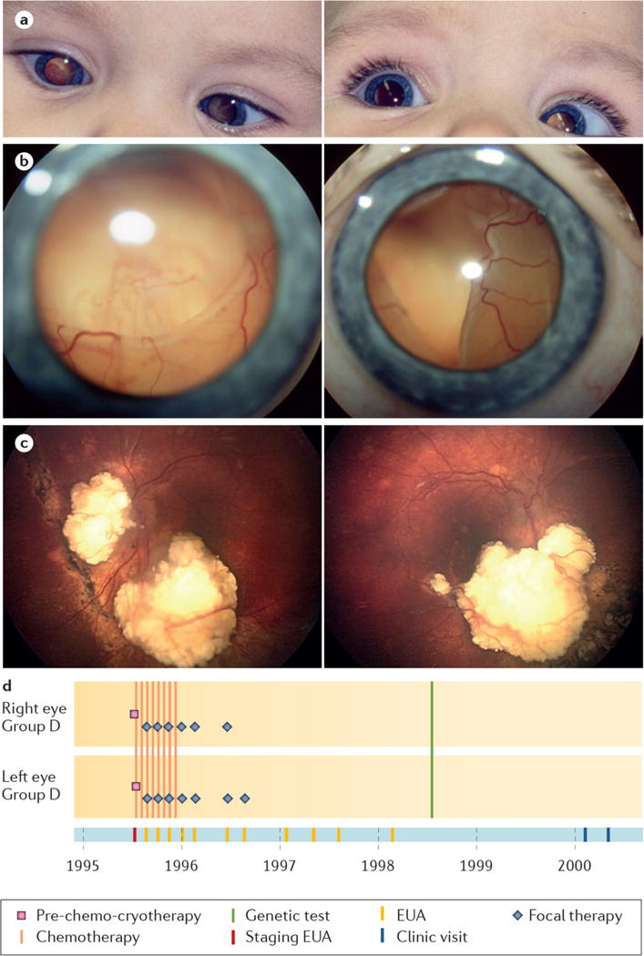
a, Detection of photoleukocoria in both eyes at age 8 months. b, Retinal photography at diagnosis showed total retinal detachment with underlying large tumours in the right (left image) and left eyes (right image). c, Both eyes have calcified tumor remnants and attached retinas 20 years after diagnosis, with 0.25 vision, right eye and 0.1 vision, left eye (Vision decimal system, 1 = normal, 0.1=legal blindness). d, eCCRB time line of shows treatment with IVC and focal therapy, then only follow-up visits.