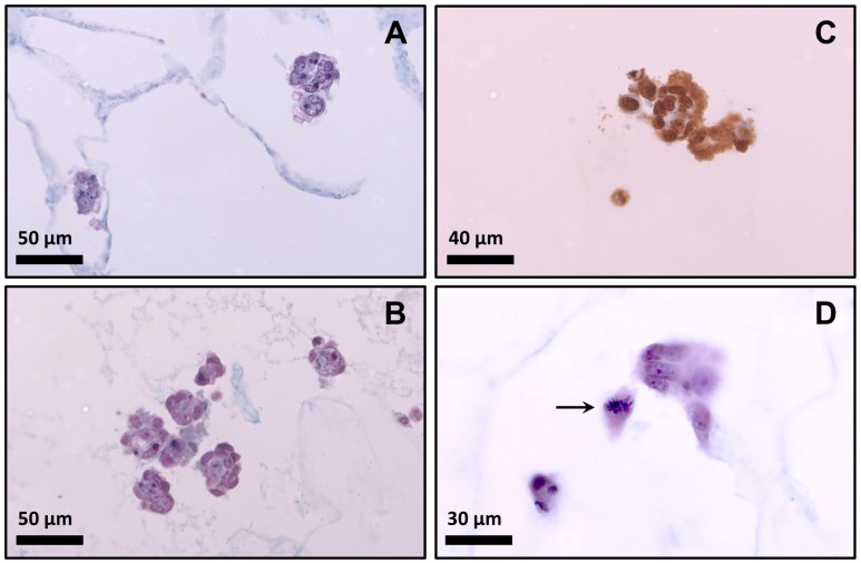
Figure 8.
Behavior of myoblasts inside the scaffold after 48 h of seeding. (A) Photomicrograph of scaffold Ag (histochemical technique); (B) Photomicrograph of scaffold Bg (histochemical technique); (C) Cells seeded on scaffold Bg showing positive immunostaining for BrdU (immuno histochemical technique); (D) Photomicrograph of scaffold Bg (arrowed cell in metaphase).

