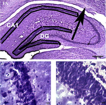
Fig. 1.

Sample microphotographs to illustrate the regions investigated. A A 2× image of a Nissl-stained section from a control rat demonstrating the CA1 area and the dentate gyrus in the right hippocampus. The arrow indicates the point of intersection to determine the CA1 area. B A 63x image of a Nissl-stained section from a control rat showing pyramidal cell bodies in the stratum pyramidale of the CA1 area, and C A 63x image of a Nissl-stained section from a control rat showing a densely packed granule cell layer of the dentate gyrus. Scale bars as indicated
