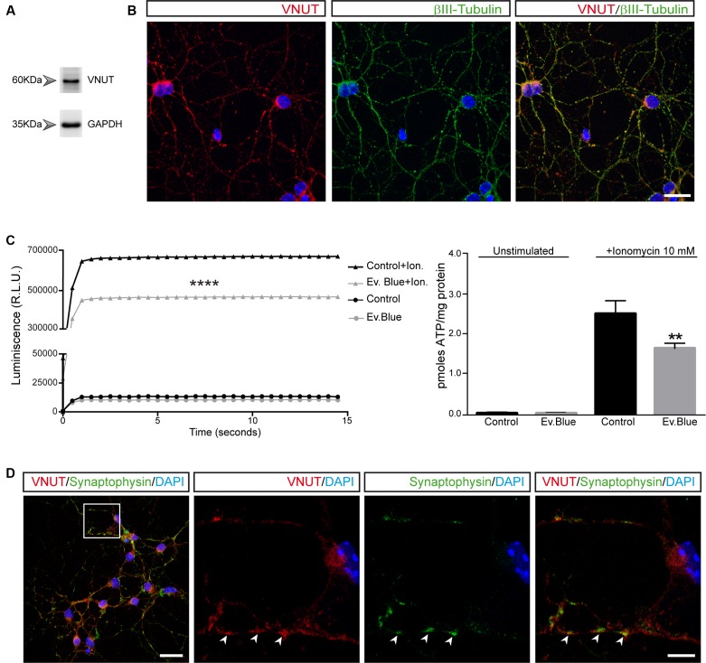
FIGURE 1.
Vesicular nucleotide transporter (VNUT) is expressed by cultured cerebellar granule cells. Identification of VNUT in granule cells by western blotting (A) and immunofluorescence (B). Cells were stained with antibodies against VNUT (red) and βIII-tubulin (green). Scale bar: 20 μm. (C) ATP release to the extracellular medium by granule cells. The graph shows the luminescence values of cells maintained in the presence or absence of Evans Blue (2 μM) and stimulated with ionomycin (10 μM) or left unstimulated. The right graph represents the pmoles of ATP released per mg of protein in the different experimental conditions. The values represent the mean ±SEM (n = 3: ∗∗p<0.01, ∗∗∗∗p<0.0001, unpaired Student’s t-test). (D) Representative confocal microscopy images of the immunostaining of granule cell neurons for VNUT (red) and synaptophysin (green). The nuclei are counterstained with DAPI (blue). Scale bar: 20 μm. The inset represents a 4x magnification of the cell indicated. The arrows indicate the most prominent VNUT and synaptophysin positive vesicles. Scale bar: 5 μm.

