
Fig. 10.
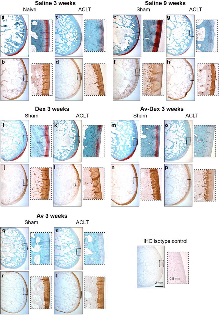
Histology and collagen II IHC of sagittal sections taken from lateral condyles following ACLT or sham surgery. Serial sections were stained for safranin O/fast green (top rows) or immunostained for type II collagen (bottom rows). Insets show higher magnification images along the articular surface of the condyles.